
If your pet shows any of these signs
Animals rarely show that their mouth hurts. By the time an owner notices, the disease has usually progressed a long way.
- Bad breath has become noticeably worse
- Stops partway through a meal, or chews on one side only
- Has suddenly started avoiding hard treats
- Drools, or dislikes having the mouth touched
- Blood shows at the gums, or the gums are red and swollen
- Swelling on the face or under the eye · a hole has appeared
- A tooth is loose or has changed colour
- Frequent sneezing or nosebleeds (this can come from an upper tooth)
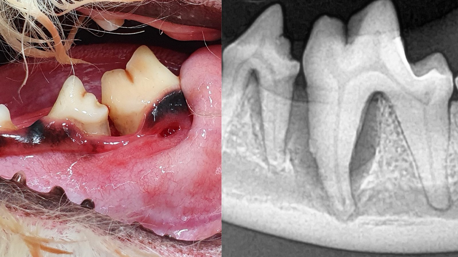
Periodontal disease
Calculus and bacterial plaque cause inflammation of the gums. When inflammation persists, the alveolar bone supporting the teeth is progressively lost. Lost bone cannot always be fully restored.
The amount of visible calculus does not necessarily reflect the severity of periodontal disease. A mouth may look relatively clean even when substantial bone loss has occurred beneath the gumline.
So before scaling we perform full-mouth dental radiography and probe the periodontal pockets. We look at the images with you and confirm which teeth can be saved and which cannot.
- What we do
- Full-mouth dental radiography · periodontal probing · scaling · root planing · periodontal surgery or extraction when required · polishing
- Why anaesthesia is necessary
- Cleaning beneath the gumline and obtaining diagnostic radiographs of each tooth require the patient to remain completely still. Anaesthesia-free scaling removes only visible calculus and does not provide comprehensive treatment for periodontal disease.
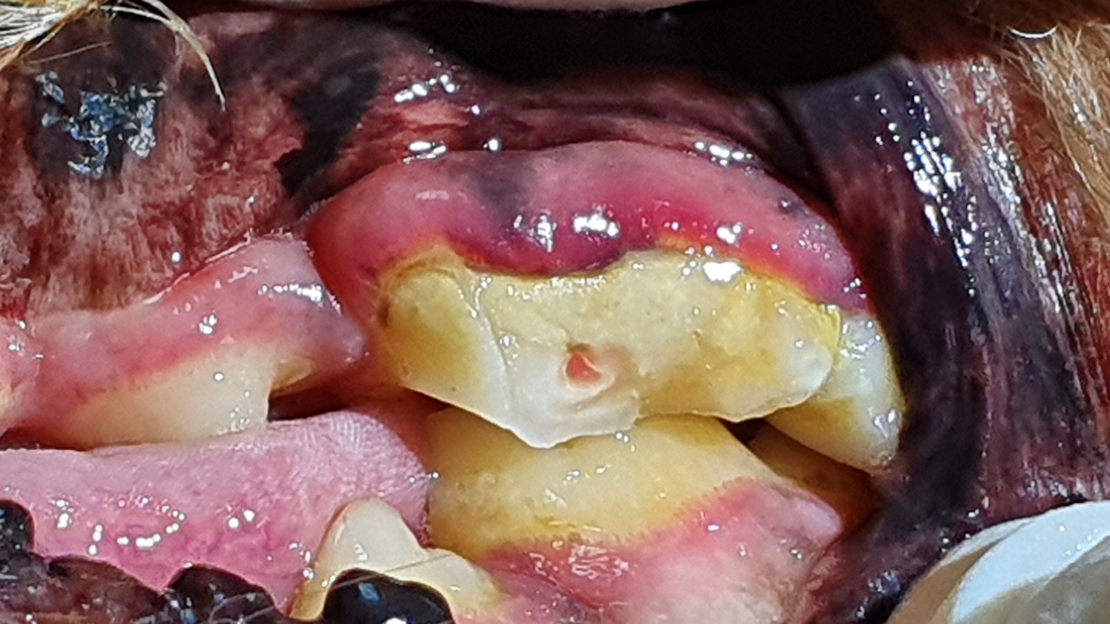
Tooth fracture
Dogs often fracture a canine or a large cheek tooth while chewing bones, antlers or hard toys. When a fracture exposes the pulp, it is painful and bacteria can enter the root canal system.
Animals often hide oral pain. Rather than “They are eating, so they must be fine,” the reality may be that they are eating despite the pain.
A fracture with exposed pulp is vulnerable to bacterial infection and, if left untreated, can lead to pulp necrosis and infection around the root tip. Prompt treatment improves the chance of preserving the tooth.
- When the tooth can be saved
- If the root and surrounding tissues remain suitable for treatment, root canal treatment followed by a crown may preserve the natural tooth.
- When the tooth cannot be saved
- Where infection has reached the root and destroyed the surrounding bone, or the root itself is split, extraction is the better choice. That judgement is made on the radiograph.
Root canal treatment · crowns
Root canal treatment removes the infected pulp, disinfects the space and fills it, saving the tooth. It works on the same principle as root canal treatment in people, adapted to animal anatomy by a veterinarian who has practised in both human and veterinary dentistry so that it can be completed in a single visit.
Afterwards the remaining tooth is protected with a crown or a composite restoration so that it does not fracture again. A composite restoration is finished on the same day; a crown requires a second anaesthetic about a week later while the crown is made.
Functionally important teeth, such as canines and large cheek teeth, are particularly valuable to preserve when possible.
After treatment, we take follow-up radiographs as appropriate to confirm that the tissues around the root remain stable. Our published cases include a two-year follow-up after treatment.
- Indications
- Fractured teeth with exposed pulp · discoloured teeth · teeth whose root can be preserved
- Fees
- Root canal, 1 canal KRW 650,000 · each additional canal KRW 165,000 · crown KRW 900,000 · composite restoration (3 surfaces) KRW 250,000 — see the fees page
Feline tooth resorption (FORL)
Tooth resorption is common in cats. Dental hard tissue is progressively lost through resorption, and exposure of the underlying dentine can be extremely painful.
From the outside, it may appear only as a small area of red gum or tissue at the tooth margin, so it is easily missed. Dental radiographs are essential for assessing the roots, classifying the lesion and planning treatment.
Treatment is either extraction or crown amputation (removing only the upper part of the tooth), depending on the form of the lesion. That decision is also made by looking at whether the root remains on the radiograph.
If your cat suddenly drops food while eating, cannot close its mouth, or drools, it needs to be checked.
- What we do
- Full-mouth radiography · lesion assessment · extraction or crown amputation · suturing
- After treatment
- Once the pain is gone, many animals show a marked change in appetite and temperament.
Periapical abscess
When infection and pus accumulate around a tooth root, a draining tract may extend through the bone and open onto the skin. Swelling or a wound below the eye can easily be mistaken for a dermatological condition.
The most common causes are a fracture of the upper carnassial tooth (fourth premolar) or severe periodontal disease.
Antibiotics may reduce the inflammation temporarily, but the problem is likely to recur unless the affected tooth is treated.
Where a problem in an upper tooth extends towards the nose, sneezing, nosebleeds and nasal discharge can appear.
- What we do
- Identify the causative tooth (radiography) · root canal treatment or extraction · drainage · closure of an oronasal fistula where needed
- Signs that are easily missed
- Swelling below the eye · a facial draining tract or wound · sneezing or nosebleeds from one nostril · recurring facial dermatitis
Extraction · oral surgery
Leaving a tooth that cannot be saved means leaving pain and infection in place. In severe periodontal disease many animals eat better and become more active after extraction.
Owners often ask, “Will my pet still be able to eat without teeth?” In practice, many animals eat better after extraction than they did with painful teeth. Chronic oral inflammation may also affect overall health.
Extraction is a surgical procedure: the gum is opened, the bone contoured, the root removed completely and the site sutured. Breaking off the root of an infected tooth and leaving it behind causes far greater problems later.
That is why a dental radiograph must always be taken after extraction to confirm that no root remains.
- What we do
- Confirm root anatomy on radiographs · raise a mucoperiosteal flap · contour the alveolar bone · remove the root completely · suture · obtain a post-extraction radiograph
- Fees
- The extent of an extraction varies greatly with the tooth, the number of roots and the state of the gum, so we advise individually at the consultation.
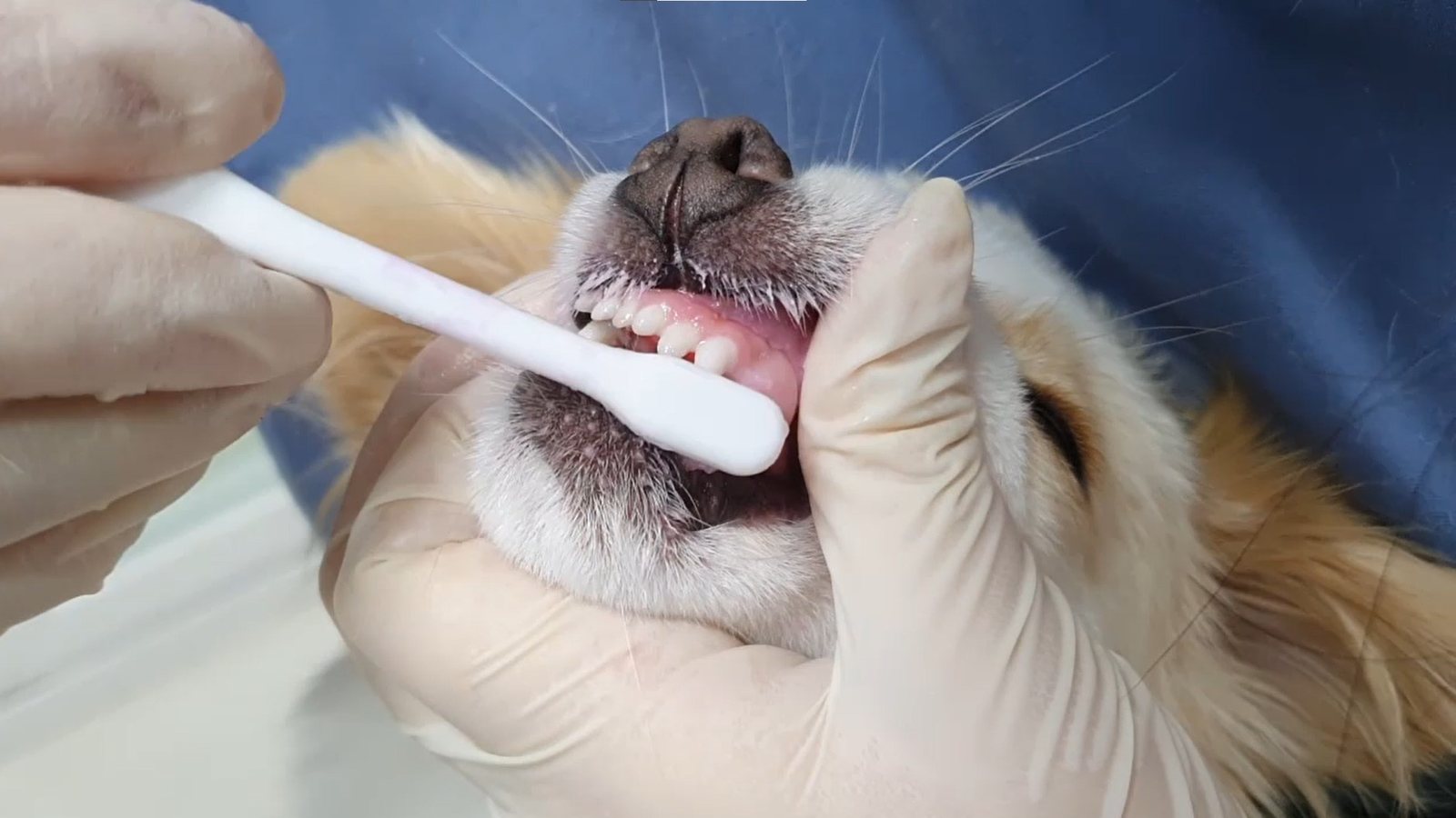
Home care after treatment
Scaling is a beginning, not an end. Calculus starts to build up again from the moment it is removed.
Brushing is by far the most effective thing you can do. Once a day is best; if that is difficult, clean along the gum line at least every other day.
Dental chews and oral products support brushing — they do not replace it. We do not recommend very hard items such as beef bones or antlers, which are a common cause of tooth fracture.
Please schedule regular oral check-ups after dental treatment. Detecting recurrent disease early may reduce the extent of future treatment and the likelihood of another anaesthetic procedure.
- Getting started with brushing
- Touch around the mouth → touch the gums → finger brush → toothbrush. Let your pet grow used to it slowly over several days.
- What to avoid
- Beef bones · antlers · hard nylon toys · ice — all common causes of tooth fracture.
How treatment proceeds
Consultation and treatment are separate appointments. If you would like both on the same day, the booking is confirmed once the deposit is paid.
We examine as much of the mouth as can be seen while your pet is awake, and explain the tests needed and the likely scope of treatment. A consultation-only booking carries no deposit.
Blood tests, chest radiographs, echocardiography and any other necessary items are performed. The anaesthetic plan, fluids and medication are adjusted according to the results.
Every tooth is radiographed and the periodontal pockets measured. This is where the actual scope of treatment is settled. At this point we call you in and go through the radiographs with you.
We carry out the agreed procedures — scaling, periodontal treatment, root canal treatment, crowns, composite restorations and extractions. You can observe the entire process through the glass.
We radiograph again to confirm the work was done properly. You may wait beside your pet for the 30 minutes of recovery.
We explain how to brush and which foods and toys to be careful of, and arrange a follow-up if one is needed.